NOCTUA
STIMULATED RAMAN IMAGING
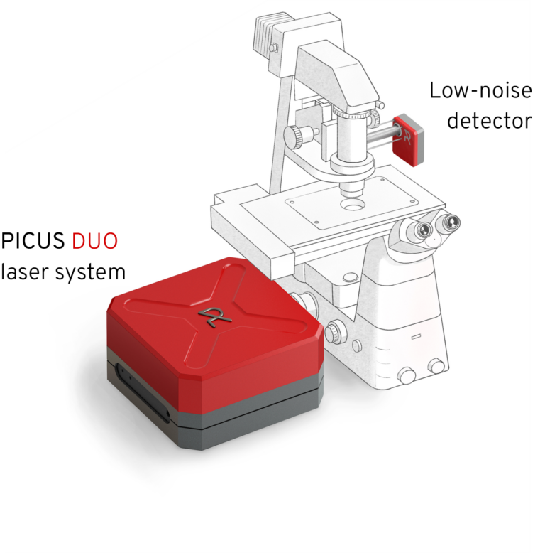
- Add-on to Evident Scientific (form. Olympus) BX63, FV3000 or Nikon Ti2
- Fully-computer controlled & adjustment-freee
- Intrinsically confocal for imaging 3D specimens
- Minimum phototoxicity by near-infrared laser
label-free imaging
with highest contrast
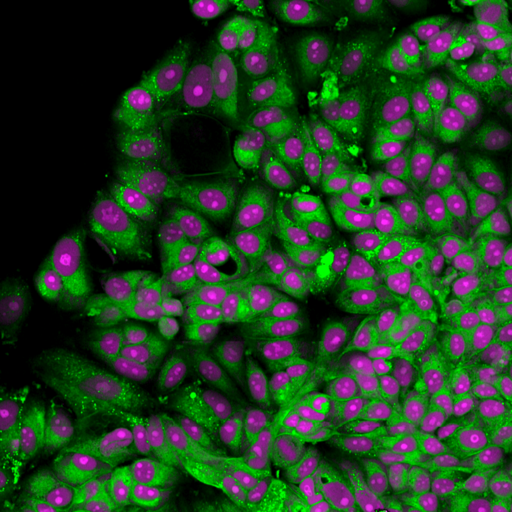
Identify nuclei & cytoplasm
without staining
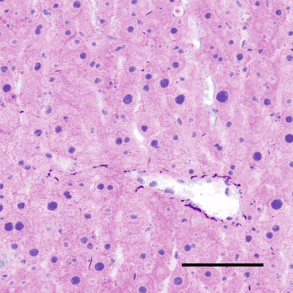
Our Noctua microscopy system allows imaging fresh and frozen tissue sections for pathological assessment without complex staining. The SRS image shows cell nuclei (purple) and cell bodies (pink) in mouse liver tissue. This label-free, virtual H&E contrast correlates well with classic H&E Staining.
Reveal & Explore
the hidden structures
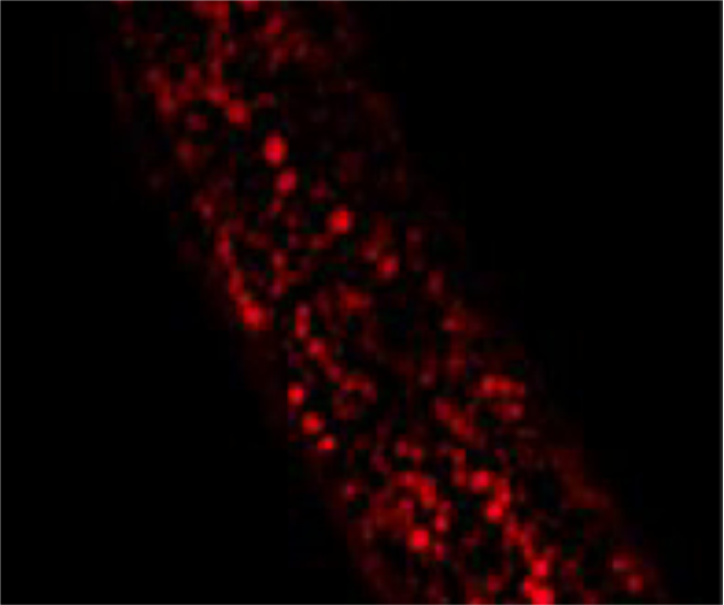
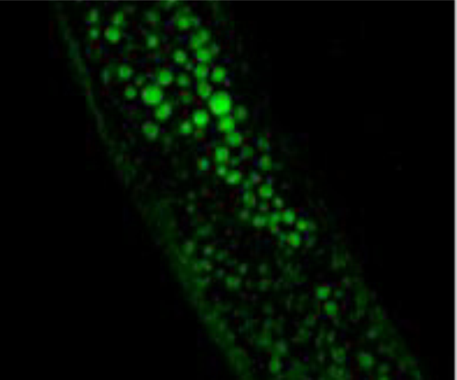
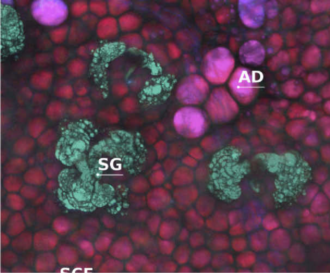
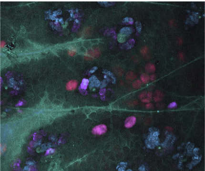
Our Noctua microscopy system allows imaging structures and events in physiological relevant conditions, that are inaccessible with fluorescent labels. The SRS images show unsaturated fatty acids and cholesterol in C.elegans (upper panel). The lower panel shows dermal structures, formed by lipids, in the skin of a mouse ear (left) and a topically applied, unlabeled drug penetrating through the skin (right).
Observe fine details
with high spectral resolution
Our NOCTUA stimulated Raman microscopy system features a low-noise spectral tuning mode to resolve sharp spectral resonances. This feature is especially helpful in the fingerprint spectral region.
- Image molecular bonds across complete Raman spectrum from 700 – 3100 cm-1
- Switch from fingerprint to CH region in milliseconds
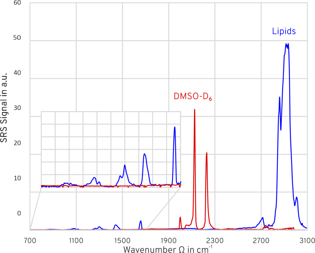
Recorded SRS spectrum of lipids (blue) and deuterated DMSO (red). Courtesy of Prof. Cheng, Boston Uni.
Balanced detector
low-noise imaging
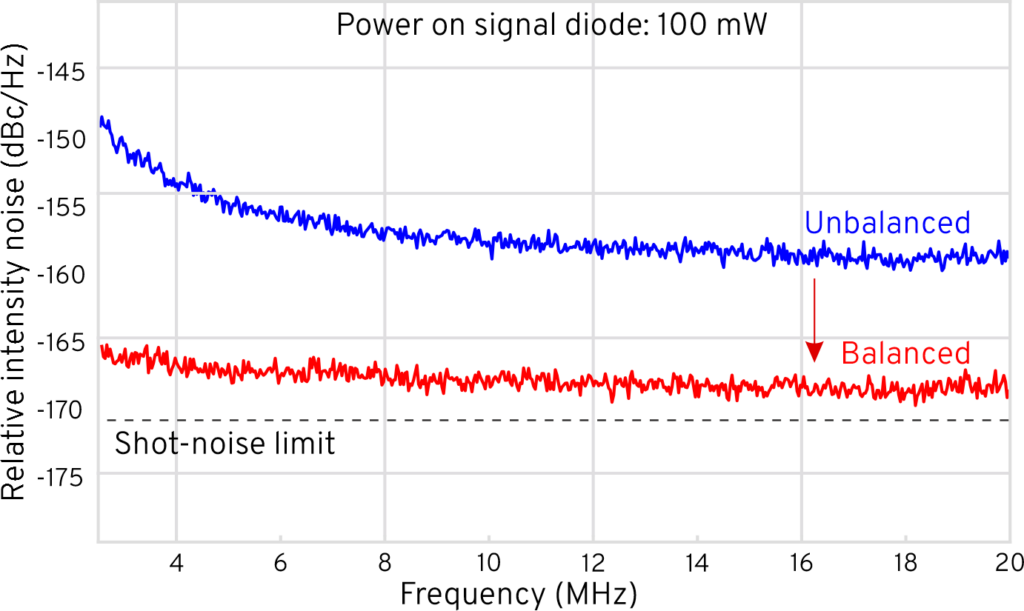
To overcome laser noise, which typically prevents imaging systems from reaching the theoretical shot-noise limit, we utilize a balanced detector. By measuring laser noise in a reference beam and sub-tracting it from the SRS signal after the sample, noise is canceled out, leading to an improved signal-to-noise ratio and increased imaging speed.
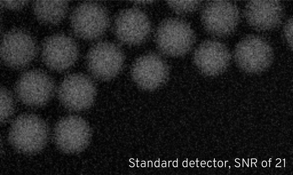
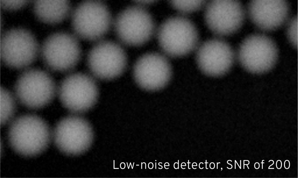
As shown below, we have achieved a noise suppression by 10 dBc with our fiber-coupled balanced detector. For illustration, the upper right-hand site shows an SRS image of plastic beads with a signal-to-noise ratio of 21 obtained with a standard detector. In contrast, the lower right-hand site shows the same image obtained with our low-noise detector with an improved SNR of 200.
highest available
tuning speed
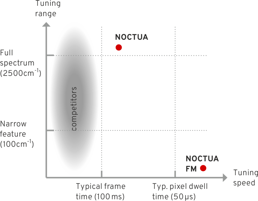
Our Noctua system offers a tuning speed of only milliseconds between 700 and 3100 cm-1. The tuning mechanism allows sweeping and switching operation and requires no external delay.
The combination of wide tunability across the full Raman spectrum and fast tunability is unmatched by any conventional system. This makes our system the predestined solution for video-rate CARS and SRS imaging at multiple wavenumbers or the rapid acquisition of hyperspectral datasets.
HYPERSPECTRAL IMAGING 10x FASTER
- Tuning between 700 – 3100 cm-1 in milliseconds
- No external delay or modulator required
- [100 spectral points, 256×256 pixels, 10 microseconds dwell time] in just 1 minute
noctua
Specifications
| Microscope specifications (typical) |
||
|---|---|---|
| Field of view | 350 μm x 350 μm | |
| Excitation objective | 25x, NA 1.1 | |
| Collection condenser | NA 0.9 | |
| Resolution | < 500 nm | |
| Pixel dwell time | > 1 μs | |
| Laser specifications | Output A | Output B |
|---|---|---|
| Tuning range | 750-980 nm | 1025 - 1052 nm |
| Tuning speed | <100 ms | <100 ms |
| Average power | 100 - 250 mW | > 300 mW |
| Pulse duration | 7 - 10 ps | 2 - 3 ps |
| Covered wavenumbers | 700 - 3100 cm-1 | |
| Spectral bandwidth | < 12 cm -1 | |
| Repetition rate | 40 MHz | |
| Mechanicel dimensions (excluding microscope stativ) |
||
|---|---|---|
| Table top laser + scanner | 42 x 65 x 25 cm3 | |
| Supply unit (off table) | 43 x 45 x 26 cm3 | |
Availability Notice:
Due to region-specific IP regulations and restrictions, Noctua is not available in all areas worldwide. Not available in the USA. We appreciate your understanding that we are currently able to serve only select regions. If you have any questions about availability in your country, please feel free to contact us.
