Stain-free and section-free histology
For cancer diagnosis
Intraoperative histology within minutes
Refined Lasers offers a novel technology for stain-free tissue diagnostics within minutes. Based on a stain-free yet molecular-specific microscope, our histology solution delivers tissue images with H&E-equivalent contrast from freshly excised, thick specimens. Without the need for time-consuming staining or sectioning, our solution allows minimizing the time required for a reliable diagnosis to just a few minutes.
- No sample preparation
- No sectioning
- No staining
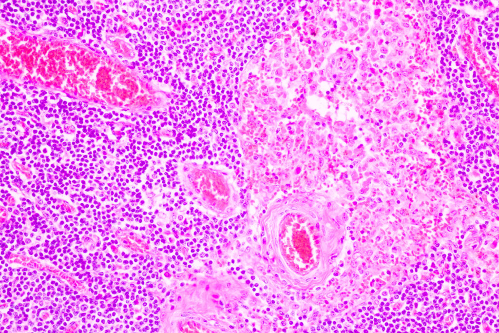
H&E image of breast cancer tissue.
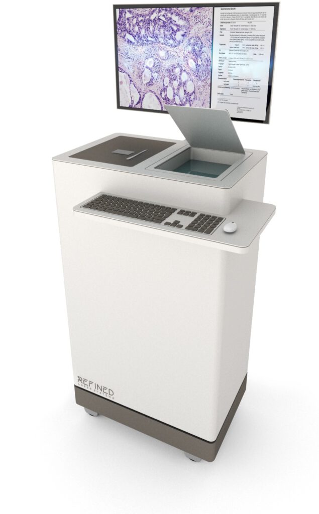
We are working on integrating our technology for use in surgery rooms and pathology labs. The fully automated systems will provide digital H&E images of whole tissue samples during surgery with maximum ease of use. Minimizing the time required to produce an H&E image will enable rapid diagnosis and feedback loops between pathologists and surgeons via a digital connection. Identifying and diagnosing cancerous tissue directly during surgery will aid the surgeon in complete and accurate removal of cancerous tissue and avoid repeat surgeries. A cloud-based and AI-powered algorithm will help pathologists make decisions under time-critical conditions.